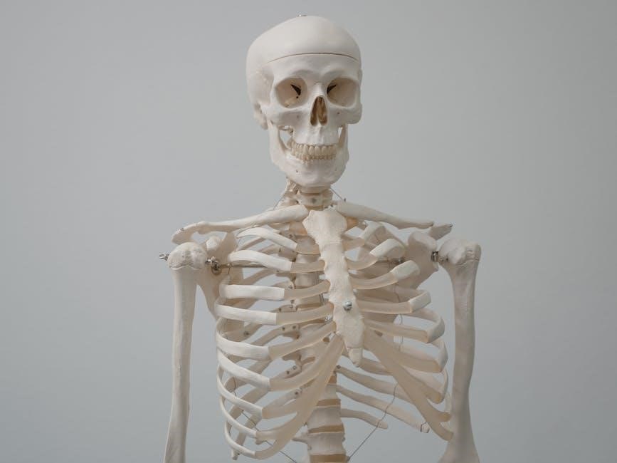
Netter Anatomy PDF is a cornerstone of medical education, offering detailed illustrations and clinical insights. Its integration of art and science makes it a vital resource for students and professionals.
Overview of Netter Anatomy PDF
The Netter Anatomy PDF is a renowned medical resource offering detailed, clinically relevant illustrations of the human body. Authored by Frank H. Netter, it provides clear, brilliant depictions from a clinician’s perspective. The atlas includes updated radiologic images, videos, and comprehensive anatomical views, making it a gold standard in anatomy education. Available in high-quality PDF format, it supports medical students and professionals with its portability and accessibility on platforms like Google Drive. Its integration of art and science has made it a widely respected and essential tool in anatomy studies.
Importance of Netter Anatomy in Medical Education
Netter Anatomy PDF is a cornerstone of medical education, providing a visual vocabulary for understanding human anatomy. Its detailed illustrations and clinical relevance bridge the gap between theoretical knowledge and practical application. Widely regarded as the gold standard, it helps students and professionals master complex anatomical concepts. The PDF format enhances portability, allowing easy access for study and reference. Its emphasis on anatomic relationships and clinically relevant views makes it indispensable for both learning and real-world application in healthcare settings.
Structure and Content of the Netter Atlas
The Netter Atlas is meticulously organized, offering comprehensive coverage of human anatomy through detailed illustrations and concise explanations. Each section focuses on specific anatomical systems, enhanced with clinically relevant views and radiologic images. The atlas includes over 550 exquisite plates, providing a clear visual understanding of anatomic relationships. Recent editions feature updates such as new illustrations, clinical tables, and integrated multimedia resources, including videos and 3D interactive models. This structured approach ensures it remains a vital tool for both medical students and practicing professionals, bridging education and clinical practice effectively.
History of Netter Anatomy Atlas
Frank H. Netter’s Atlas of Human Anatomy, first published in 1989, revolutionized medical education with its precise illustrations. Netter’s artistic brilliance and clinical expertise laid the foundation for this iconic resource, now translated into 16 languages and widely regarded as a cornerstone of anatomical learning.
Frank H. Netter and His Contributions to Anatomy
Frank H. Netter, MD, was a visionary physician and artist whose detailed anatomical illustrations transformed medical education. His unique ability to merge clinical accuracy with artistic precision created visually compelling and educational content. Netter’s work emphasized anatomic relationships and clinical relevance, making complex anatomy accessible to students and professionals alike. His contributions laid the foundation for modern anatomical education, earning his atlas a reputation as the gold standard in the field. Netter’s legacy continues to inspire learning and advancements in anatomy.
Evolution of the Netter Atlas Over Editions
The Netter Atlas has undergone significant enhancements over its editions, incorporating advancements in medical knowledge and visual technology. The 7th edition introduced updated illustrations by Dr. Carlos A. G. Machado and expanded radiologic imaging. Later editions added 3D interactive anatomy and online modules, providing immersive learning experiences. Each update reflects a commitment to accuracy and innovation, ensuring the atlas remains a cutting-edge resource for anatomy education. These improvements have solidified its reputation as an indispensable tool for medical professionals and students.
Impact of Netter’s Work on Modern Anatomy Education
Frank Netter’s work has revolutionized anatomy education by providing visually precise and clinically relevant illustrations. His atlas has set a standard for understanding human anatomy, bridging the gap between artistic representation and scientific accuracy. The integration of radiologic images and 3D interactive tools in recent editions has enhanced learning, making complex anatomical concepts more accessible. Netter’s contributions have influenced generations of medical professionals, solidifying his role as a pioneer in modern anatomy education and beyond.

Key Features of Netter Anatomy PDF
Netter Anatomy PDF offers detailed, clinically relevant illustrations, comprehensive anatomical coverage, and integrated radiologic images. It combines artistic precision with scientific accuracy, enhancing learning and retention for students and professionals.
Detailed Illustrations and Their Significance
Netter Anatomy PDF is renowned for its exquisite, detailed illustrations that provide clear, brilliant depictions of the human body. Created by Dr. Frank H. Netter, these illustrations emphasize anatomic relationships and clinically relevant views, making them indispensable for medical education. The artwork combines artistic precision with scientific accuracy, offering a visual vocabulary that enhances understanding and retention. These illustrations are particularly valuable for clinicians, as they highlight structures and connections most pertinent to diagnosis and treatment. The inclusion of radiologic images and videos further enriches the learning experience, bridging anatomy with real-world clinical applications.
Clinical Relevance in Anatomy Education
Netter Anatomy PDF excels in linking anatomical knowledge to clinical practice, making it a gold standard for medical education. Its focus on anatomic relationships and clinically relevant views ensures that students and professionals can apply the information directly to patient care. The atlas emphasizes structures and connections most pertinent to diagnosis and treatment, providing a clinician-oriented perspective. This practical approach helps bridge the gap between theoretical learning and real-world medical scenarios, enhancing the ability to understand and address clinical challenges effectively.
Comprehensive Coverage of Human Anatomy
Netter Anatomy PDF provides an extensive and detailed exploration of human anatomy, covering all major body systems and structures. Its illustrations and descriptions are meticulously organized to ensure a thorough understanding of anatomical relationships. From skeletal and muscular systems to nervous and circulatory systems, the atlas offers a complete visual and textual guide. Cross-sectional views, radiologic images, and anatomical variations further enhance its comprehensiveness, making it an indispensable resource for mastering human anatomy in depth.
Updates in Recent Editions
Recent editions of Netter Anatomy PDF feature updated illustrations by Dr. Frank H. Netter and Dr. Carlos A. G. Machado, along with new radiologic images and clinical tables. The inclusion of 3D interactive anatomy content and videos from Netter’s Online Dissection Modules enhances learning. These updates ensure the atlas remains current with modern anatomical knowledge and educational needs, providing students and professionals with the latest tools for understanding human anatomy in a clinically relevant context.
Target Audience
Medical students, healthcare professionals, and researchers rely on Netter Anatomy PDF for its detailed visuals and practical applications. Artists and anatomical illustrators also benefit from its precise depictions.
Medical Students and Healthcare Professionals
Netter Anatomy PDF is indispensable for medical students and healthcare professionals, offering detailed illustrations and clinically relevant content. Its clear depictions of anatomic relationships aid in understanding complex structures, making it a valuable tool for both study and practice. The PDF format enhances portability, allowing access on various devices. This resource bridges the gap between theoretical knowledge and practical application, ensuring professionals can quickly reference anatomical details in clinical settings. Its precision and clarity make it an essential asset for medical education and ongoing professional development.
Artists and Anatomical Illustrators
Netter Anatomy PDF is a vital resource for artists and anatomical illustrators, offering meticulous and lifelike depictions of human anatomy. Its detailed illustrations provide inspiration and reference for creating accurate and visually compelling artwork. The clarity and precision of Netter’s work aid artists in understanding anatomical relationships, enabling them to capture the human form with greater accuracy. This resource is particularly valued for its ability to bridge art and science, making it an indispensable tool for both creative and educational purposes in the field of anatomical drawing.
Researchers and Academics
Netter Anatomy PDF is an invaluable resource for researchers and academics, providing a comprehensive and detailed visual reference for human anatomy. Its precise illustrations and clinical correlations make it a cornerstone for scholarly work. The inclusion of radiologic images and updated content ensures it remains a cutting-edge tool for anatomical research. Additionally, the availability of 3D interactive modules and online resources enhances its utility for academic studies and presentations, making it a go-to resource for both advanced research and educational purposes in the field of anatomy.

Unique Selling Points
Netter Anatomy PDF stands out for its artistic precision, clinical relevance, and integration of radiologic images. Its 3D interactive tools and digital accessibility make it indispensable for modern learning.
Artistic and Scientific Precision in Illustrations
Netter Anatomy PDF is renowned for its meticulous blend of artistic and scientific accuracy. Frank H. Netter’s illustrations are celebrated for their lifelike detail, capturing anatomical structures with unmatched clarity. Each drawing combines aesthetic appeal with precise scientific information, making complex anatomy accessible. The vibrant, high-quality images emphasize clinically relevant views, aiding both medical education and practice. Netter’s work bridges art and science, offering a visual vocabulary that enhances understanding and retention. The illustrations’ precision ensures they remain a gold standard in anatomy education, while updates like 3D tools and radiologic images further enrich their educational value.
Practical Applications of Anatomical Knowledge
Netter Anatomy PDF equips users with practical anatomical insights essential for clinical practice. Its detailed illustrations guide clinicians in diagnosis, treatment planning, and surgical approaches. Radiologic images enhance understanding of anatomical structures in real-world scenarios. The atlas bridges theory and application, aiding medical professionals in visualizing complex systems. For educators, Netter’s visuals simplify teaching, while students benefit from clear, clinically relevant depictions. The integration of 3D tools further supports practical learning, making it an indispensable resource for both education and professional practice in medicine and related fields.
Integration of Radiologic Images and Videos
Netter Anatomy PDF enhances learning with integrated radiologic images and videos, providing a multidimensional understanding of anatomy. The inclusion of 3D interactive anatomy and online dissection modules allows users to visualize complex structures dynamically. Radiologic images, such as X-rays and MRIs, correlate anatomical illustrations with real-world clinical scenarios. These multimedia resources bridge the gap between theoretical knowledge and practical application, making the atlas invaluable for both students and professionals in medicine and related fields.
Learning Resources
Netter Anatomy PDF is complemented by 3D Interactive Anatomy, online dissection modules, and flash cards. These tools enhance interactive learning, making complex anatomy engaging and accessible for students and professionals.
Netter’s 3D Interactive Anatomy
Netter’s 3D Interactive Anatomy offers a dynamic learning experience with interactive modules. Students can explore 3D views of anatomical structures, enhancing their understanding of complex relationships. The tool provides detailed visuals, allowing users to rotate, zoom, and layer tissues. It includes videos and quizzes to reinforce learning. This resource is particularly beneficial for visual learners, offering a deeper engagement with anatomy. Accessible online, it complements the PDF atlas, making it an indispensable tool for medical students and professionals seeking to master human anatomy effectively. Its interactive nature bridges the gap between textbook learning and real-world application.
Online Dissection Modules
Online Dissection Modules provide a virtual platform for exploring human anatomy through realistic simulations. These modules replicate actual dissection experiences, offering detailed visuals and step-by-step guidance. They enable students to examine structures layer by layer, enhancing comprehension of spatial relationships. The modules are integrated with Netter’s illustrations, ensuring consistency and depth. This resource is invaluable for medical students, allowing them to practice and review anatomy in a flexible, accessible manner. The interactive nature of these modules bridges traditional dissection with modern digital learning tools, fostering a deeper understanding of human anatomy.
Flash Cards for Quick Revision
Netter’s Flash Cards are an excellent tool for rapid review and memorization of key anatomical structures; Featuring high-quality illustrations from Frank H. Netter, MD, these cards provide concise, clinically relevant information. Designed for portable study, they allow learners to quickly reinforce their understanding of complex anatomical concepts. The flash cards are particularly useful for exam preparation, offering a straightforward way to test knowledge and identify areas needing further review. This resource complements the Netter Atlas, making it a valuable addition to any anatomy study routine.

Practical Applications
Netter Anatomy PDF is widely used in medical education, clinical practice, and artistic endeavors, bridging anatomy with real-world applications, enhancing learning, and supporting professional development across diverse fields.
Use in Medical Curriculum and Exams
Netter Anatomy PDF is a cornerstone in medical education, serving as a primary resource for anatomy courses. Its detailed illustrations and clinical correlations make it indispensable for exam preparation, particularly for USMLE and other medical licensing exams. The PDF format enhances portability, allowing students to study anywhere. The atlas is often integrated into curricula for its clear depiction of anatomic relationships, aiding in both theoretical understanding and practical application. Its relevance extends to clinical training, where it bridges the gap between classroom learning and real-world patient care.
Application in Clinical Practice
Netter Anatomy PDF is invaluable in clinical practice, providing clear, detailed visuals that aid healthcare professionals in diagnosing and treating patients. Its illustrations highlight anatomical relationships crucial for surgical planning and understanding complex medical conditions. Clinicians rely on Netter’s work to visualize structures before procedures, enhancing precision and decision-making. The inclusion of radiologic images further supports real-world applications, making it a trusted tool for bridging theoretical knowledge with practical patient care in various medical specialties.
Role in Art and Anatomical Drawing
Netter Anatomy PDF has inspired artists and anatomical illustrators by blending scientific accuracy with artistic mastery. Its detailed, lifelike illustrations provide a visual foundation for understanding human anatomy, making it a valuable resource for figure drawing and anatomical art. Artists use Netter’s work to study muscle structure, joint mechanics, and proportional accuracy, enabling them to create realistic and anatomically correct depictions of the human form. The PDF format enhances accessibility, allowing artists to reference these illustrations effortlessly on digital devices, fostering creativity and precision in their work.
Digital Access and Benefits
Netter Anatomy PDF offers enhanced learning through digital convenience, portability, and cost-effectiveness. It provides anytime access to detailed illustrations, 3D interactive anatomy, and additional online modules.
Advantages of the PDF Format
The Netter Anatomy PDF offers unparalleled portability and accessibility, allowing users to access detailed anatomical illustrations anytime, anywhere. Its digital format enables easy searching, bookmarking, and hyperlink navigation, enhancing study efficiency. The PDF is cost-effective compared to physical copies and reduces the need for bulky textbooks. Additionally, it supports environmental sustainability by minimizing paper usage. The format also integrates seamlessly with devices like tablets and laptops, making it ideal for modern learners. Its clarity and functionality make it a preferred choice for both students and professionals in the medical field.
Accessibility and Portability
The Netter Anatomy PDF is designed for easy access across various devices, ensuring that users can study on-the-go. Its portability allows learners to carry a comprehensive anatomy resource without the bulk of physical textbooks. The digital format is accessible on smartphones, tablets, and laptops, making it convenient for both in-class and remote learning. This flexibility supports diverse learning styles and environments, empowering users to review anatomical details anytime, anywhere, and enhancing overall study efficiency and engagement with the material.
Cost-Effectiveness of Digital Versions
Digital versions of Netter Anatomy PDF offer significant cost savings compared to physical copies. The PDF format eliminates the need for bulky textbooks, reducing expenses while maintaining comprehensive content. Affordable pricing makes it accessible to students and professionals without compromising on quality. Additionally, digital access often includes free updates and supplementary materials like 3D models and videos, enhancing the learning experience. This cost-effective solution ensures users gain maximum value without financial strain, making it a practical choice for modern learners;
Comparison with Other Anatomy Resources
Netter Anatomy PDF stands out for its detailed, clinically relevant illustrations and comprehensive coverage, making it a preferred choice over other anatomy resources for students and professionals.
Netter Atlas vs. Gray’s Anatomy
While both Netter’s Atlas and Gray’s Anatomy are esteemed anatomy resources, they cater to different learning styles. Netter’s Atlas is renowned for its visually oriented, clinically relevant illustrations, making it a favorite among medical professionals. Gray’s Anatomy, while comprehensive and historically significant, is more text-heavy and detailed. Netter’s focus on anatomic relationships and practical applications gives it an edge for clinicians, whereas Gray’s remains a thorough, encyclopedic reference. Both are invaluable, but Netter’s artistic precision often makes it preferred for visual learners and clinical practice.
Netter Atlas vs. Other Anatomy Atlases
Netter’s Atlas stands out among anatomy resources due to its unique blend of artistic precision and clinical relevance. Unlike other atlases, Netter’s illustrations are created by a physician-artist, ensuring accuracy and a focus on anatomic relationships crucial for clinical practice. While other atlases may prioritize detailed descriptions, Netter’s emphasizes visual learning, making it a preferred choice for medical professionals. Its updates, including radiologic images and 3D interactive tools, further enhance its utility, distinguishing it as a modern, comprehensive resource in anatomy education.
Unique Strengths of Netter’s Work
Netter’s Atlas excels due to its exceptional artistry and scientific accuracy, blending the precision of a physician with the flair of an artist. Frank Netter’s illustrations provide a clear, lifelike representation of anatomy, aiding both students and professionals. Unlike other resources, Netter’s work emphasizes anatomic relationships and clinical relevance, making it a gold standard in medical education. Its detailed yet concise approach ensures that complex anatomical concepts are presented in an accessible, visually engaging manner, solidifying its reputation as an indispensable tool for understanding human anatomy.
Downloading and Accessing Netter Anatomy PDF
Accessing Netter Anatomy PDF is straightforward via platforms like Google Drive or official medical websites, offering high-quality formats for easy learning and reference on various devices.
Legal and Ethical Considerations
Downloading Netter Anatomy PDF requires adherence to copyright laws and ethical standards. Ensure you obtain the file from authorized sources or purchase it legally to avoid piracy. Unauthorized distribution violates intellectual property rights and undermines the work of authors and publishers. Always prioritize ethical access to support the creators and maintain academic integrity. Many institutions provide legitimate access through subscriptions or libraries, ensuring compliance with legal guidelines while benefiting from this invaluable resource.
Steps to Access the PDF Version
To access the Netter Anatomy PDF, visit official platforms like Google Drive or the publisher’s website. Purchase or subscribe to gain legal access. Ensure the source is reputable to avoid unauthorized versions. Many academic institutions provide access through their libraries or online portals. Additionally, platforms like Amazon or eBookstores offer digital versions for download. Always verify the authenticity of the source to ensure a high-quality, complete edition of the atlas.
Platforms for Downloading the Atlas
Popular platforms for downloading the Netter Anatomy PDF include Amazon, Google Drive, and the publisher’s official website. Additionally, many academic institutions offer access through their libraries or online portals. Digital stores like eBooks.com and VitalSource also provide secure options for purchasing and downloading. Always ensure the platform is reputable to avoid unauthorized copies. Some platforms may require a subscription or one-time purchase for access to the full PDF version of the atlas.

Legacy and Impact
Frank H. Netter’s work remains the gold standard in medical illustration, inspiring generations with its precision and clarity, shaping anatomy education and clinical practice worldwide.
Frank H. Netter’s Legacy in Anatomy
Frank H. Netter’s legacy is defined by his transformative contributions to medical illustration. His detailed, lifelike anatomical art has educated generations of healthcare professionals, blending scientific accuracy with artistic brilliance. Netter’s work, particularly his iconic atlas, has become a cornerstone of anatomy education, renowned for its clarity and clinical relevance. His illustrations emphasize anatomic relationships crucial for diagnosis and treatment, solidifying his impact on both medical practice and education. Netter’s legacy continues to inspire, ensuring his work remains indispensable in the field of anatomy.
Continued Relevance in Modern Education
Netter Anatomy PDF remains a cornerstone in modern medical education due to its unmatched clarity and clinical relevance. Its detailed illustrations and concise explanations continue to bridge the gap between theoretical knowledge and practical application. The integration of radiologic images and digital tools enhances its accessibility and portability, making it a preferred resource for today’s learners. Despite advancements in technology, Netter’s work retains its value, ensuring it remains a vital tool for students and professionals alike in understanding human anatomy.
Future of Netter Anatomy Resources
The future of Netter Anatomy resources lies in advancing digital integration and interactive learning. With the rise of AI and VR, these tools are expected to enhance anatomical education further. The incorporation of 3D models, real-time simulations, and personalized learning platforms will make Netter’s work even more accessible and engaging. Additionally, continuous updates with the latest medical discoveries will ensure the content remains relevant. The legacy of Netter’s work will continue to evolve, adapting to technological advancements while maintaining its core commitment to excellence in anatomy education.
Netter Anatomy PDF remains the gold standard for anatomy education, combining artistic excellence with scientific precision. It is an indispensable resource for learners and professionals alike.
Final Thoughts on Netter Anatomy PDF
Netter Anatomy PDF is a timeless resource, blending artistic precision with scientific accuracy. Its detailed illustrations and clinical relevance make it essential for medical students and professionals. The integration of 3D models and radiologic images enhances learning. Portable and cost-effective, it remains a cornerstone in anatomy education, bridging traditional and modern learning methods. Highly recommended for anyone seeking a comprehensive understanding of human anatomy, Netter’s work continues to inspire and educate future generations in the medical field.
Recommendations for Potential Users
Netter Anatomy PDF is highly recommended for medical students, healthcare professionals, and anatomy enthusiasts. Its detailed illustrations and clinical relevance make it ideal for both learning and reference. For artists, the anatomical accuracy provides exceptional guidance. Researchers and educators will appreciate its comprehensive coverage. Available in digital formats, it offers portability and accessibility, making it a versatile tool for modern learners. This resource is indispensable for anyone seeking a deep understanding of human anatomy, blending tradition with innovative learning aids.
